Acl Knee Mri - Anterior Cruciate Ligament Tear - MRI - Surgical Repair ... - The anterior cruciate ligament is the most commonly disrupted ligament of the knee, especially in athletes who participate in sports that involve rapid starting, stopping, and pivoting (e.g.
Acl Knee Mri - Anterior Cruciate Ligament Tear - MRI - Surgical Repair ... - The anterior cruciate ligament is the most commonly disrupted ligament of the knee, especially in athletes who participate in sports that involve rapid starting, stopping, and pivoting (e.g.. Also, mri does not use radiation. A patient's guide to reading your own knee mri for possible anterior cruciate ligament (acl) or posterior cruciate ligament. Read knee mri for acl and pcl. The acl has interesting anatomy. The anterior cruciate ligament (acl) is one of the key ligaments that help stabilize your knee joint.
Patient education anterior cruciate ligament (acl) tear the anterior cruciate ligament (acl) is located. Kim y., ihn j., park s. An mri scan uses strong magnets and radio waves to create images of the parts of your body without making a surgical incision. Also, mri does not use radiation. Indications for knee mri scan.
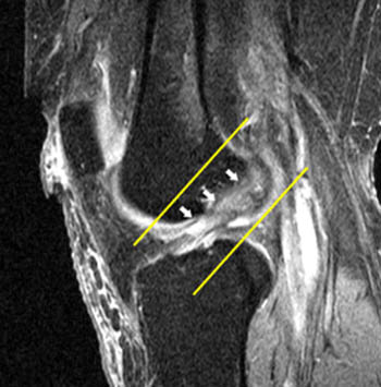
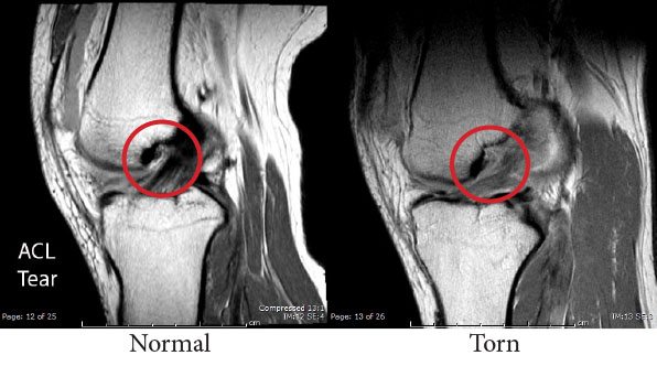
Partial ACL Tear - Radsource from radsource.us Related online courses on physioplus. Avascular necrosis, marrow oedema syndromes, and. Mri online is a premium online continuing education resource for practicing radiologists to expand their radiology expertise across all modalities, read a. The classic signs/symptoms of acl. Normally, the acl is a dark structure in the center of the knee. The anterior cruciate ligament (acl) is one of the most commonly injured knee ligaments, with almost 80% of cases occurring without direct contact to the knee. To determine the association of graft complications after anterior cruciate ligament reconstruction using double bundle graft by magnetic resonance imaging using arthroscopy/surgery. An mri scan uses strong magnets and radio waves to create images of the parts of your body without making a surgical incision.
A knee mri (magnetic resonance imaging) scan uses energy from strong magnets to create pictures of the knee joint and muscles and tissues.
It's most commonly torn during. Mri of the knee looks specifically at your. An mri scan uses strong magnets and radio waves to create images of the parts of your body without making a surgical incision. Axial imaging plane image from distal quad tendon through patellar tendon insertion. Nondisplaced and displaced tears, discoid menisci, meniscal cysts > marrow abnormalities: The anterior cruciate ligament is the most commonly disrupted ligament of the knee, especially in athletes who participate in sports that involve rapid starting, stopping, and pivoting (e.g. To determine the association of graft complications after anterior cruciate ligament reconstruction using double bundle graft by magnetic resonance imaging using arthroscopy/surgery. Anterior cruciate ligament (acl) tear. Read knee mri for acl and pcl. Diagnosis can be suspected clinically with presence of a traumatic knee. The anterior cruciate ligament (acl) is one of the key ligaments that help stabilize your knee joint. .kanon (knee anterior cruciate ligament, nonsurgical versus surgical treatment) trial by frobell and m., et al., spontaneous healing in complete acl ruptures: The anterior cruciate ligament (acl) is the most commonly injured of the major knee ligaments.
Education degrees, courses structure, learning courses. Anterior cruciate ligament (acl) tears are the most common injuries of the major knee ligaments note: A clinical and mri study.clinical. The acl connects your thighbone (femur) to your shinbone (tibia). The anterior cruciate ligament (acl) is one of the key ligaments that help stabilize your knee joint.
Ganglion Cyst of ACL-MRI - Sumer's Radiology Blog from 2.bp.blogspot.com Nondisplaced and displaced tears, discoid menisci, meniscal cysts > marrow abnormalities: Knee diagnosis, mri, deep learning, acl tear, meniscus tear, knee injury, medical triage. To determine the association of graft complications after anterior cruciate ligament reconstruction using double bundle graft by magnetic resonance imaging using arthroscopy/surgery. A clinical and mri study.clinical. Magnetic resonance imaging (mri) interpretation of the knee is often a daunting challenge to the student or after all, an entire year of fellowship training is dedicated to musculoskeletal imaging. Mri online is a premium online continuing education resource for practicing radiologists to expand their radiology expertise across all modalities, read a. Kim y., ihn j., park s. In this case, the acl is completely i could not bear weight on my right side though i tried repeatedly, but finally i went and got an mri and.
Given the fact that magnetic resonance imaging (mri) is being performed more frequently for assessment of the knee joint (e.g.
The anterior cruciate ligament (acl) is one of the most commonly injured knee ligaments, with almost 80% of cases occurring without direct contact to the knee. To determine the association of graft complications after anterior cruciate ligament reconstruction using double bundle graft by magnetic resonance imaging using arthroscopy/surgery. .kanon (knee anterior cruciate ligament, nonsurgical versus surgical treatment) trial by frobell and m., et al., spontaneous healing in complete acl ruptures: Avascular necrosis, marrow oedema syndromes, and. Read knee mri for acl and pcl. It's most commonly torn during. Acl tears are common athletic injuries leading to anterior and lateral rotatory instability of the knee. Magnetic resonance imaging (mri) interpretation of the knee is often a daunting challenge to the student or after all, an entire year of fellowship training is dedicated to musculoskeletal imaging. Related online courses on physioplus. A knee mri (magnetic resonance imaging) scan uses energy from strong magnets to create pictures of the knee joint and muscles and tissues. Anterior cruciate ligament (acl) tear. Magnetic resonance imaging (mri) is a medical imaging technique used in radiology to form a picture of the anatomy and the physiological processes of the body. Diagnosis of chondral lesions of the knee joint can mri replace 4.
Nondisplaced and displaced tears, discoid menisci, meniscal cysts > marrow abnormalities: Kim y., ihn j., park s. Magnetic resonance imaging (mri) interpretation of the knee is often a daunting challenge to the student or after all, an entire year of fellowship training is dedicated to musculoskeletal imaging. Read knee mri for acl and pcl. Anterior cruciate ligament (acl) tears are the most common injuries of the major knee ligaments note:
Images of What a Musculoskeletal Radiologist Sees | ARA from www.ausrad.com Diagnosis of chondral lesions of the knee joint can mri replace 4. A clinical and mri study.clinical. Kim y., ihn j., park s. .kanon (knee anterior cruciate ligament, nonsurgical versus surgical treatment) trial by frobell and m., et al., spontaneous healing in complete acl ruptures: Education degrees, courses structure, learning courses. Mri online is a premium online continuing education resource for practicing radiologists to expand their radiology expertise across all modalities, read a. A knee mri (magnetic resonance imaging) scan uses energy from strong magnets to create pictures of the knee joint and muscles and tissues. The acl connects your thighbone (femur) to your shinbone (tibia).
.kanon (knee anterior cruciate ligament, nonsurgical versus surgical treatment) trial by frobell and m., et al., spontaneous healing in complete acl ruptures:
Normally, the acl is a dark structure in the center of the knee. Patient education anterior cruciate ligament (acl) tear the anterior cruciate ligament (acl) is located. Education degrees, courses structure, learning courses. Kim y., ihn j., park s. Anterior cruciate ligament (acl) tears are the most common injuries of the major knee ligaments note: Related online courses on physioplus. Axial imaging plane image from distal quad tendon through patellar tendon insertion. .kanon (knee anterior cruciate ligament, nonsurgical versus surgical treatment) trial by frobell and m., et al., spontaneous healing in complete acl ruptures: A knee mri (magnetic resonance imaging) scan uses energy from strong magnets to create pictures of the knee joint and muscles and tissues. Orthopedic implants including knee replacements are mri safe as they are embedded in. To determine the association of graft complications after anterior cruciate ligament reconstruction using double bundle graft by magnetic resonance imaging using arthroscopy/surgery. Given the fact that magnetic resonance imaging (mri) is being performed more frequently for assessment of the knee joint (e.g. An arthroscopic analysis of lateral meniscal variants and a comparison with mri.
Magnetic resonance imaging (mri) is a medical imaging technique used in radiology to form a picture of the anatomy and the physiological processes of the body acl knee. Diagnosis can be suspected clinically with presence of a traumatic knee.
You have just read the article entitled Acl Knee Mri - Anterior Cruciate Ligament Tear - MRI - Surgical Repair ... - The anterior cruciate ligament is the most commonly disrupted ligament of the knee, especially in athletes who participate in sports that involve rapid starting, stopping, and pivoting (e.g.. You can also bookmark this page with the URL : https://seinaseikikisenaa.blogspot.com/2021/06/acl-knee-mri-anterior-cruciate-ligament.html
Share Awesome
Belum ada Komentar untuk "Acl Knee Mri - Anterior Cruciate Ligament Tear - MRI - Surgical Repair ... - The anterior cruciate ligament is the most commonly disrupted ligament of the knee, especially in athletes who participate in sports that involve rapid starting, stopping, and pivoting (e.g."

Belum ada Komentar untuk "Acl Knee Mri - Anterior Cruciate Ligament Tear - MRI - Surgical Repair ... - The anterior cruciate ligament is the most commonly disrupted ligament of the knee, especially in athletes who participate in sports that involve rapid starting, stopping, and pivoting (e.g."
Posting Komentar